|
Western blot analysis of 1 HEK293 2 SW480 3 HEPG2 4 MCF-7 5 mouse brain 6 Rat brain 7 Hela 8 A549 lysates, primary antibody was diluted at 1:5000, 4° over night, secondary antibody was diluted at 1:10000, 37° 1hour. |
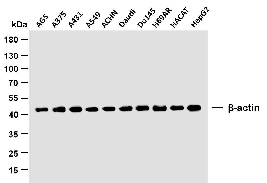 |
Various whole cell lysates were separated by 10% SDS-PAGE, and the membrane was blotted with anti-β-actin antibody. The HRP-conjugated Goat anti-Mouse IgG(H + L) antibody was used to detect the antibody. Lane 1: AGS Lane 2: A375 Lane 3: A431 Lane 4: A549 Lane 5: ACHN Lane 6: Daudi Lane 7: Du145 Lane 8: H69AR Lane 9: HACAT Lane 10: HepG2 |
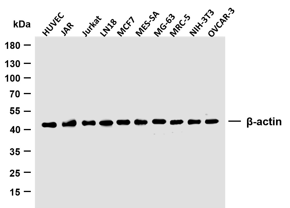 |
Various whole cell lysates were separated by 10% SDS-PAGE, and the membrane was blotted with anti-β-actin antibody. The HRP-conjugated Goat anti-Mouse IgG(H + L) antibody was used to detect the antibody. Lane 1: HUVEC Lane 2:JAR Lane 3:Jurkat Lane 4:LN18 Lane 5:MCF7 Lane 6:MES-SA Lane 7:MG-63 Lane 8:MRC-5 Lane 9:NIH-3T3 Lane 10:OVCAR-3 |
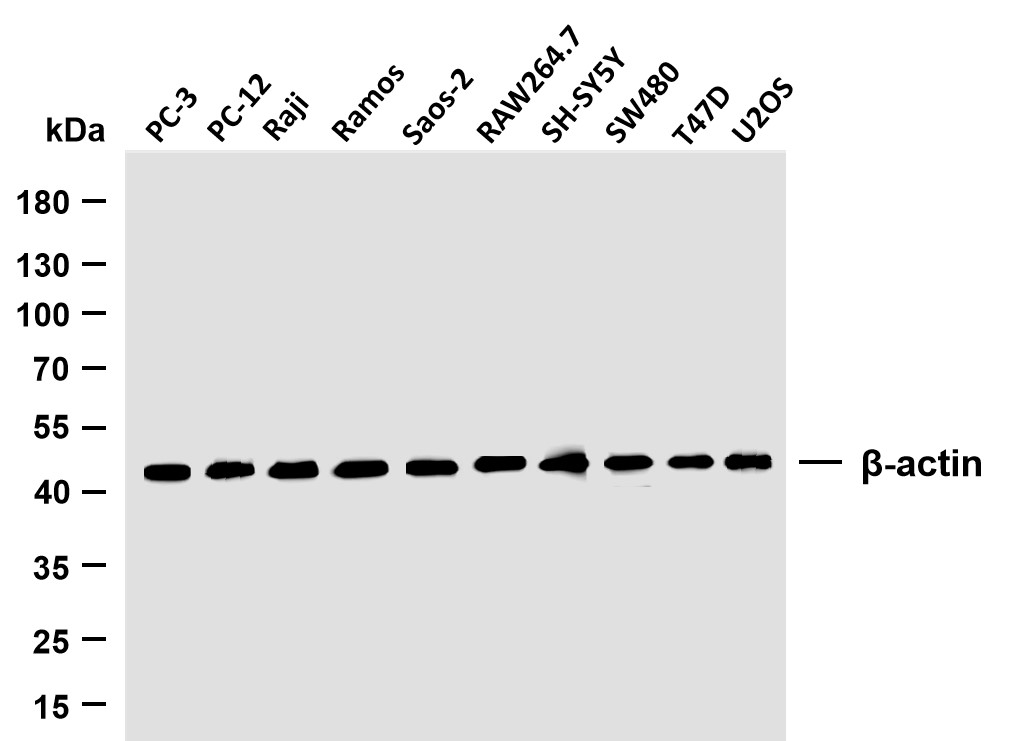 |
Various whole cell lysates were separated by 10% SDS-PAGE, and the membrane was blotted with anti-β-actin antibody. The HRP-conjugated Goat anti-Mouse IgG(H + L) antibody was used to detect the antibody. Lane 1: PC-3 Lane 2: PC-12 Lane 3: Raji Lane 4: Ramos Lane 5: Saos-2 Lane 6: RAW264.7 Lane 7: SH-SY5Y Lane 8: SW480 Lane 9: T47D Lane 10: U2OS |
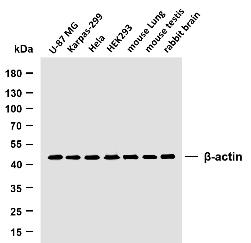 |
Various whole cell lysates were separated by 10% SDS-PAGE, and the membrane was blotted with anti-β-actin antibody. The HRP-conjugated Goat anti-Mouse IgG(H + L) antibody was used to detect the antibody. Lane 1: U-87 MG Lane 2: Karpas-299 Lane 3: Hela Lane 4: HEK293 Lane 5: Mouse Lung Lane 6: mouse testis Lane 7: Rabbit brain |


 收藏
收藏

 电话咨询
电话咨询
 在线咨询
在线咨询
 QQ
QQ
 二维码
二维码
 扫码二维码
扫码二维码