GFAP(5C8)Mouse Monoclonal Antibody

货号: BD-PM3059
收藏
规格 价格 货期 数量
  • 50μl
    1100.00
    现货
    100μl
    2000.00
    现货

产品基本信息

  • 产品货号 BD-PM3059
  • 别名 GFAP; Glial fibrillary acidic protein; GFAP
  • 产品名称 GFAP(5C8)Mouse Monoclonal Antibody
  • 类别 抗体产品
  • 基因名称 GFAP
  • 蛋白名称 Glial fibrillary acidic protein
  • Clonality Monoclonal
  • 推荐应用 WB,IHC-P,IF-P,IF-F,IF-ICC
  • 反应种属 Human,Rat,Mouse
  • 存储缓冲液 PBS, pH 7.4, containing 0.5%BSA, 0.02% New type preservative N as Preservative and 50% Glycerol.
  • Human Gene ID 2670
  • Human Gene Link http://www.ncbi.nlm.nih.gov/sites/entrez?db=gene&term=2670
  • Human Swissprot No. P14136
  • Human Swissprot Link http://www.uniprot.org/uniprotkb/P14136/entry
  • Mouse Gene ID 14580
  • Mouse Gene Link http://www.ncbi.nlm.nih.gov/sites/entrez?db=gene&term=14580
  • Mouse Swissprot No. P03995
  • Mouse Swissprot Link http://www.uniprot.org/uniprot/P03995
  • Rat Gene ID 24387
  • Rat Gene Link http://www.ncbi.nlm.nih.gov/sites/entrez?db=gene&term=24387
  • Rat Swissprot No. P47819
  • Rat Swissprot Link http://www.uniprot.org/uniprot/P47819
  • 免疫原 Synthetic Peptide of GFAP
  • 特异性 The antibody detects endogenous GFAP proteins.
  • 稀释度 WB 1:2000-5000, IF-P/IF-F/IF-ICC 1:200, IHC-P 1:50-300
  • 预测分子量 45kDa
  • 运输及保存条件 -20°C/1 year
  • 宿主 Mouse
  • 背景介绍 This gene encodes one of the major intermediate filament proteins of mature astrocytes. It is used as a marker to distinguish astrocytes from other glial cells during development. Mutations in this gene cause Alexander disease, a rare disorder of astrocytes in the central nervous system. Alternative splicing results in multiple transcript variants encoding distinct isoforms. [provided by RefSeq, Oct 2008],
  • 组织表达 Expressed in cells lacking fibronectin.
  • 细胞定位 Cytoplasm . Associated with intermediate filaments. .
  • 功能 alternative products:Isoforms differ in the C-terminal region which is encoded by alternative exons,disease:Defects in GFAP are a cause of Alexander disease (ALEXD) [MIM:203450]. Alexander disease is a rare disorder of the central nervous system. It is a progressive leukoencephalopathy whose hallmark is the widespread accumulation of Rosenthal fibers which are cytoplasmic inclusions in astrocytes. The most common form affects infants and young children, and is characterized by progressive failure of central myelination, usually leading to death usually within the first decade. Infants with Alexander disease develop a leukoencephalopathy with macrocephaly, seizures, and psychomotor retardation. Patients with juvenile or adult forms typically experience ataxia, bulbar signs and spasticity, and a more slowly progressive course.,function:GFAP, a class-III intermediate filament, is a cell-specific marker that, during the development of the central nervous system, distinguishes astrocytes from other glial cells.,online information:GFAP entry,similarity:Belongs to the intermediate filament family.,subcellular location:Associated with intermediate filaments.,subunit:Interacts with SYNM (By similarity). Isoform 3 interacts with PSEN1 (via N-terminus).,tissue specificity:Expressed in cells lacking fibronectin.,
  • 纯化 The antibody was affinity-purified from mouse ascites by affinity-chromatography using specific immunogen.

图片

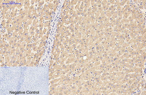
Immunohistochemical analysis of paraffin-embedded Human-liver tissue. 1,GFAP Monoclonal Antibody(5C8) was diluted at 1:200(4°C,overnight). 2, Sodium citrate pH 6.0 was used for antibody retrieval(>98°C,20min). 3,Secondary antibody was diluted at 1:200(room tempeRature, 30min). Negative control was used by secondary antibody only.
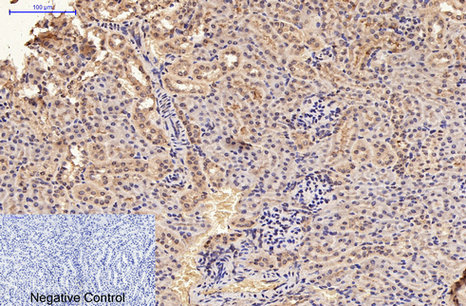
Immunohistochemical analysis of paraffin-embedded Mouse-kidney tissue. 1,GFAP Monoclonal Antibody(5C8) was diluted at 1:200(4°C,overnight). 2, Sodium citrate pH 6.0 was used for antibody retrieval(>98°C,20min). 3,Secondary antibody was diluted at 1:200(room tempeRature, 30min). Negative control was used by secondary antibody only.
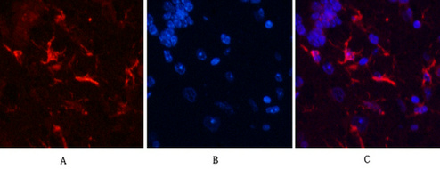
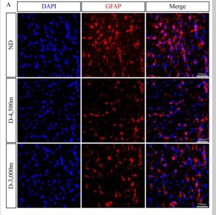
Immunofluorescence analysis of Mouse-brain tissue. 1,GFAP Monoclonal Antibody(5C8)(red) was diluted at 1:200(4°C,overnight). 2, Cy3 labled Secondary antibody was diluted at 1:300(room temperature, 50min).3, Picture B: DAPI(blue) 10min. Picture A:Target. Picture B: DAPI. Picture C: merge of A+B
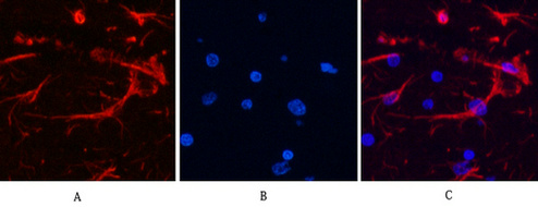
Immunofluorescence analysis of Rat-brain tissue. 1,GFAP Monoclonal Antibody(5C8)(red) was diluted at 1:200(4°C,overnight). 2, Cy3 labled Secondary antibody was diluted at 1:300(room temperature, 50min).3, Picture B: DAPI(blue) 10min. Picture A:Target. Picture B: DAPI. Picture C: merge of A+B
Immunofluorescence analysis
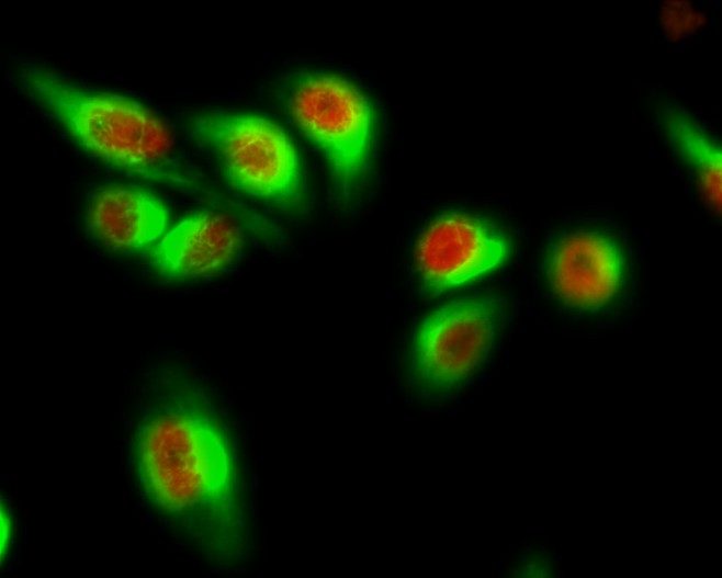
Immunofluorescence analysis of Hela cell. 1,AR Polyclonal Antibody(red) was diluted at 1:200(4° overnight). GFAP Monoclonal Antibody(green) was diluted at 1:200(4° overnight). 2, Goat Anti Rabbit Alexa Fluor 594 was diluted at 1:1000(room temperature, 50min). Goat Anti Mouse Alexa Fluor 488 was diluted at 1:1000(room temperature, 50min).
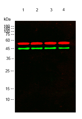
Western blot analysis of lysates from 1) Rat Brain Tissue, 2)HeLa , 3)A431, 4) PC12 cells, (Green) primary antibody was diluted at 1:1000, 4°over night, secondary antibody was diluted at 1:10000, 37° 1hour. (Red) Tubulin β Polyclonal Antibody antibody was diluted at 1:5000 as loading control, 4° over night,secondary antibody was diluted at 1:10000, 37° 1hour.

产品详情

文献引用 (0)

产品问答

内容已折叠

相关产品

内容已折叠

最新浏览的产品

评论内容:
上传图片:
商品评价:
提交评论
« »