AR Rabbit Polyclonal Antibody

货号: BD-PT5538
收藏
规格 价格 货期 数量
  • 50μl
    1100.00
    现货
    100μl
    2000.00
    现货

产品基本信息

  • 产品货号 BD-PT5538
  • 别名 AR; DHTR; NR3C4; Androgen receptor; Dihydrotestosterone receptor; Nuclear receptor subfamily 3 group C member 4
  • 产品名称 AR Rabbit Polyclonal Antibody
  • 类别 抗体产品
  • 基因名称 AR
  • 蛋白名称 Androgen receptor
  • Clonality Polyclonal
  • 推荐应用 IF-P,IF-F,IF-ICC,WB,IHC-P,ELISA
  • 反应种属 Human,Mouse,Rat
  • 浓度 1 mg/ml
  • 存储缓冲液 Liquid in PBS containing 50% glycerol, 0.5% BSA and 0.02% New type preservative N.
  • Human Gene ID 367
  • Human Gene Link http://www.ncbi.nlm.nih.gov/sites/entrez?db=gene&term=367
  • Human Swissprot No. P10275
  • Human Swissprot Link http://www.uniprot.org/uniprotkb/P10275/entry
  • Mouse Gene ID 11835
  • Mouse Gene Link http://www.ncbi.nlm.nih.gov/sites/entrez?db=gene&term=11835
  • Mouse Swissprot No. P19091
  • Mouse Swissprot Link http://www.uniprot.org/uniprot/P19091
  • Rat Gene ID 24208
  • Rat Gene Link http://www.ncbi.nlm.nih.gov/sites/entrez?db=gene&term=24208
  • Rat Swissprot No. P15207
  • Rat Swissprot Link http://www.uniprot.org/uniprot/P15207
  • 免疫原 Synthesized peptide derived from the Internal region of human AR.
  • 特异性 AR Polyclonal Antibody detects endogenous levels of AR protein.
  • 稀释度 IF-P/IF-F/IF-ICC 1:50-200, WB 1:500-2000, ELISA 1:10000-20000, IHC-P 1:50-300
  • 预测分子量 99kDa
  • 运输及保存条件 -20°C/1 year
  • 宿主 Rabbit
  • 同种型 Rabbit,IgG
  • 背景介绍 The androgen receptor gene is more than 90 kb long and codes for a protein that has 3 major functional domains: the N-terminal domain, DNA-binding domain, and androgen-binding domain. The protein functions as a steroid-hormone activated transcription factor. Upon binding the hormone ligand, the receptor dissociates from accessory proteins, translocates into the nucleus, dimerizes, and then stimulates transcription of androgen responsive genes. This gene contains 2 polymorphic trinucleotide repeat segments that encode polyglutamine and polyglycine tracts in the N-terminal transactivation domain of its protein. Expansion of the polyglutamine tract from the normal 9-34 repeats to the pathogenic 38-62 repeats causes spinal bulbar muscular atrophy (Kennedy disease). Mutations in this gene are also associated with complete androgen insensitivity (CAIS). Two alternatively spliced variants encoding distinct isoform
  • 组织表达 [Isoform 2]: Mainly expressed in heart and skeletal muscle. ; [Isoform 3]: Expressed in basal and stromal cells of the prostate (at protein level).
  • 细胞定位 Nucleus . Cytoplasm . Detected at the promoter of target genes (PubMed:25091737). Predominantly cytoplasmic in unligated form but translocates to the nucleus upon ligand-binding. Can also translocate to the nucleus in unligated form in the presence of RACK1. .
  • 信号通路 Oocyte meiosis;Pathways in cancer;Prostate cancer;
  • 功能 disease:Defects in AR are the cause of androgen insensitivity syndrome (AIS) [MIM:300068]; previously known as testicular feminization syndrome (TFM). AIS is an X-linked recessive form of pseudohermaphroditism due end-organ resistance to androgen. Affected males have female external genitalia, female breast development, blind vagina, absent uterus and female adnexa, and abdominal or inguinal testes, despite a normal 46,XY karyotype.,disease:Defects in AR are the cause of androgen insensitivity syndrome partial (PAIS) [MIM:312300]; also known as Reifenstein syndrome. PAIS is characterized by hypospadias, hypogonadism, gynecomastia, genital ambiguity, normal XY karyotype, and a pedigree pattern consistent with X-linked recessive inheritance. Some patients present azoospermia or severe oligospermia without other clinical manifestations.,disease:Defects in AR are the cause of spinal and bulbar muscular atrophy X-linked type 1 (SMAX1) [MIM:313200]; also known as Kennedy disease. SMAX1 is an X-linked recessive form of spinal muscular atrophy. Spinal muscular atrophy refers to a group of neuromuscular disorders characterized by degeneration of the anterior horn cells of the spinal cord, leading to symmetrical muscle weakness and atrophy. SMAX1 occurs only in men. Age at onset is usually in the third to fifth decade of life, but earlier involvement has been reported. It is characterized by slowly progressive limb and bulbar muscle weakness with fasciculations, muscle atrophy, and gynecomastia. The disorder is clinically similar to classic forms of autosomal spinal muscular atrophy. SMAX1 is caused by trinucleotide CAG repeat expansion. In SMAX1 patients the number of Gln ranges from 38 to 62. Longer expansions result in earlier onset and more severe clinical manifestations of the disease.,disease:Defects in AR may play a role in metastatic prostate cancer. The mutated receptor stimulates prostate growth and metastases development despite of androgen ablation. This treatment can reduce primary and metastatic lesions probably by inducing apoptosis of tumor cells when they express the wild-type receptor.,domain:Composed of three domains: a modulating N-terminal domain, a DNA-binding domain and a C-terminal steroid-binding domain. In the presence of bound steroid the ligand-binding domain interacts with the N-terminal modulating domain, and thereby activates AR transcription factor activity. Agonist binding is required for dimerization and binding to target DNA. The transcription factor activity of the complex formed by ligand-activated AR and DNA is modulated by interactions with coactivator and corepressor proteins. Interaction with RANBP9 is mediated by both the N-terminal domain and the DNA-binding domain. Interaction with EFCAB6/DJBP is mediated by the DNA-binding domain.,function:Steroid hormone receptors are ligand-activated transcription factors that regulate eukaryotic gene expression and affect cellular proliferation and differentiation in target tissues. Transcription factor activity is modulated by bound coactivator and corepressor proteins. Transcription activation is down-regulated by NR0B2. Activated, but not phosphorylated, by HIPK3.,miscellaneous:In the absence of ligand, steroid hormone receptors are thought to be weakly associated with nuclear components; hormone binding greatly increases receptor affinity. The hormone-receptor complex appears to recognize discrete DNA sequences upstream of transcriptional start sites.,miscellaneous:The level of tyrosine phosphorylation may serve as a diagnostic tool to predict patient outcome in response to hormone-ablation therapy. Inhibition of tyrosine phosphorylation may be an effective intervention target for hormone-refractory prostate cancer.,miscellaneous:Transcriptional activity is enhanced by binding to RANBP9.,online information:Androgen receptor entry,polymorphism:The poly-Gln region of AR is highly polymorphic and the number of Gln varies in the population (from 17 to 26). A smaller size of the poly-Gln region may be associated with the development of prostate cancer.,polymorphism:The poly-Gly region of AR is polymorphic and ranges from 24 to 31 Gly. A poly-Gly region shorter or equal to 23 may be associated with the development of androgenetic alopecia.,PTM:Phosphorylated in prostate cancer cells in response to several growth factors including EGF. Phosphorylation is induced by c-Src kinase (CSK). Tyr-534 is one of the major phosphorylation sites and an increase in phosphorylation and Src kinase activity is associated with prostate cancer progression.,PTM:Sumoylated on Lys-386 (major) and Lys-520.,similarity:Belongs to the nuclear hormone receptor family.,similarity:Belongs to the nuclear hormone receptor family. NR3 subfamily.,similarity:Contains 1 nuclear receptor DNA-binding domain.,subunit:Binds DNA as a homodimer. Part of a ternary complex containing AR, EFCAB6/DJBP and PARK7. Interacts with HIPK3 and NR0B2 in the presence of androgen. The ligand binding domain interacts with MYST2/HBO1 in the presence of dihydrotestosterone. Interacts with EFCAB6/DJBP, PELP1, PQBP1, RANBP9, RBAK, SPDEF, SRA1, TGFB1I1, ZNF318 and RREB1. Interacts with ZMIZ1/ZIMP10 and ZMIZ2/ZMIP7 which both enhance its transactivation activity. Interacts with SLC30A9 and RAD54L2/ARIP4 (By similarity). Interacts via the ligand-binding domain with LXXLL and FXXLF motifs from NCOA1, NCOA2, NCOA3, NCOA4 and MAGEA11. The AR N-terminal poly-Gln region binds Ran resulting in enhancement of AR-mediated transactivation. Ran-binding decreases as the poly-Gln length increases. Interacts with HIP1 (via coiled coil domain).,tissue specificity:Isoform 2 is mainly expressed in heart and skeletal muscle.,
  • 纯化 The antibody was affinity-purified from rabbit antiserum by affinity-chromatography using epitope-specific immunogen.

图片

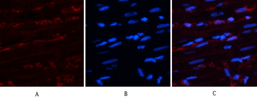
Immunofluorescence analysis of rat-heart tissue. 1,AR Polyclonal Antibody(red) was diluted at 1:200(4°C,overnight). 2, Cy3 labled Secondary antibody was diluted at 1:300(room temperature, 50min).3, Picture B: DAPI(blue) 10min. Picture A:Target. Picture B: DAPI. Picture C: merge of A+B
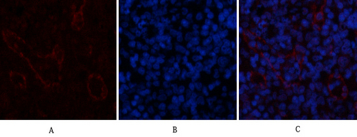
Immunofluorescence analysis of rat-spleen tissue. 1,AR Polyclonal Antibody(red) was diluted at 1:200(4°C,overnight). 2, Cy3 labled Secondary antibody was diluted at 1:300(room temperature, 50min).3, Picture B: DAPI(blue) 10min. Picture A:Target. Picture B: DAPI. Picture C: merge of A+B
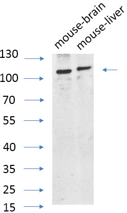
Western Blot analysis of various cells using primary antibody diluted at 1:1000(4°C overnight). Secondary antibody:Goat Anti-rabbit IgG IRDye 800( diluted at 1:5000, 25°C, 1 hour). Cell lysate was extracted by Minute™ Plasma Membrane Protein Isolation and Cell Fractionation Kit(SM-005, Inventbiotech,MN,USA).
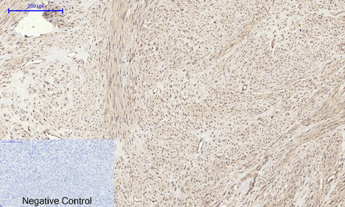
Immunohistochemical analysis of paraffin-embedded Human-uterus tissue. 1,AR Polyclonal Antibody was diluted at 1:200(4°C,overnight). 2, Sodium citrate pH 6.0 was used for antibody retrieval(>98°C,20min). 3,Secondary antibody was diluted at 1:200(room tempeRature, 30min). Negative control was used by secondary antibody only.
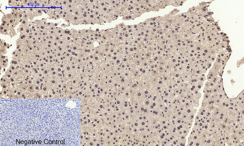
Immunohistochemical analysis of paraffin-embedded Rat-heart tissue. 1,AR Polyclonal Antibody was diluted at 1:200(4°C,overnight). 2, Sodium citrate pH 6.0 was used for antibody retrieval(>98°C,20min). 3,Secondary antibody was diluted at 1:200(room tempeRature, 30min). Negative control was used by secondary antibody only.
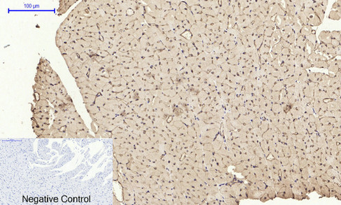
Immunohistochemical analysis of paraffin-embedded Mouse-liver tissue. 1,AR Polyclonal Antibody was diluted at 1:200(4°C,overnight). 2, Sodium citrate pH 6.0 was used for antibody retrieval(>98°C,20min). 3,Secondary antibody was diluted at 1:200(room tempeRature, 30min). Negative control was used by secondary antibody only.
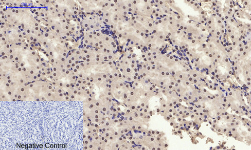
Immunohistochemical analysis of paraffin-embedded Mouse-kidney tissue. 1,AR Polyclonal Antibody was diluted at 1:200(4°C,overnight). 2, Sodium citrate pH 6.0 was used for antibody retrieval(>98°C,20min). 3,Secondary antibody was diluted at 1:200(room tempeRature, 30min). Negative control was used by secondary antibody only.
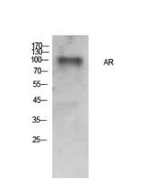
Western Blot analysis of Hela cells using AR Polyclonal Antibody.. Secondary antibody was diluted at 1:20000
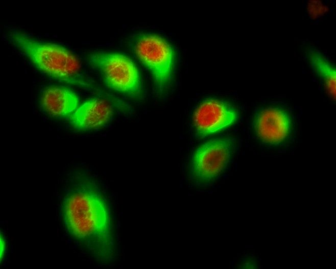
Immunofluorescence analysis of Hela cell. 1,AR Polyclonal Antibody(red) was diluted at 1:200(4° overnight). GFAP Monoclonal Antibody(green) was diluted at 1:200(4° overnight). 2, Goat Anti Rabbit Alexa Fluor 594 was diluted at 1:1000(room temperature, 50min). Goat Anti Mouse Alexa Fluor 488 was diluted at 1:1000(room temperature, 50min).

产品详情

文献引用 (0)

产品问答

内容已折叠

相关产品

内容已折叠

最新浏览的产品

评论内容:
上传图片:
商品评价:
提交评论
« »