 |
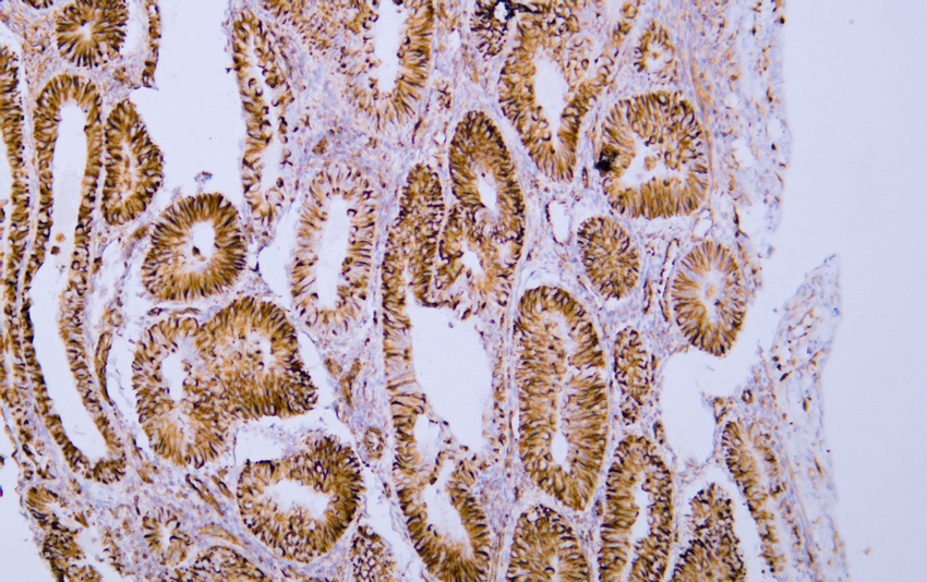
Human endometrial adenocarcinoma tissue was stained with Anti-Vimentin Antibody |
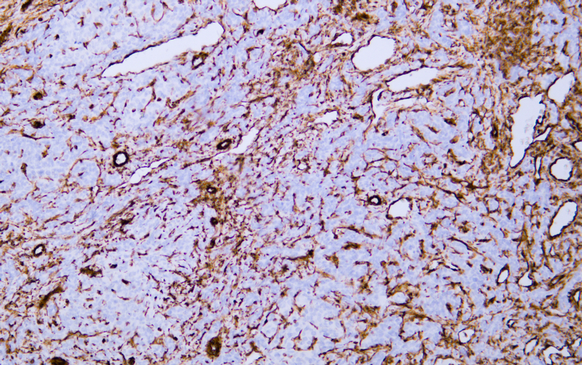 |
Human hepatocellular carcinoma tissue was stained with Anti-Vimentin Antibody |
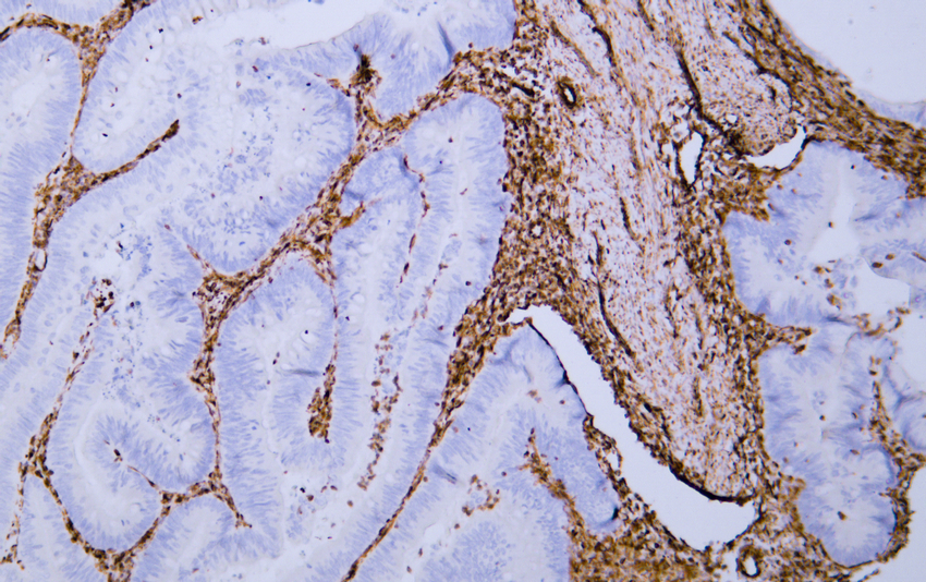 |
Human rectal carcinoma tissue was stained with Anti-Vimentin Antibody |
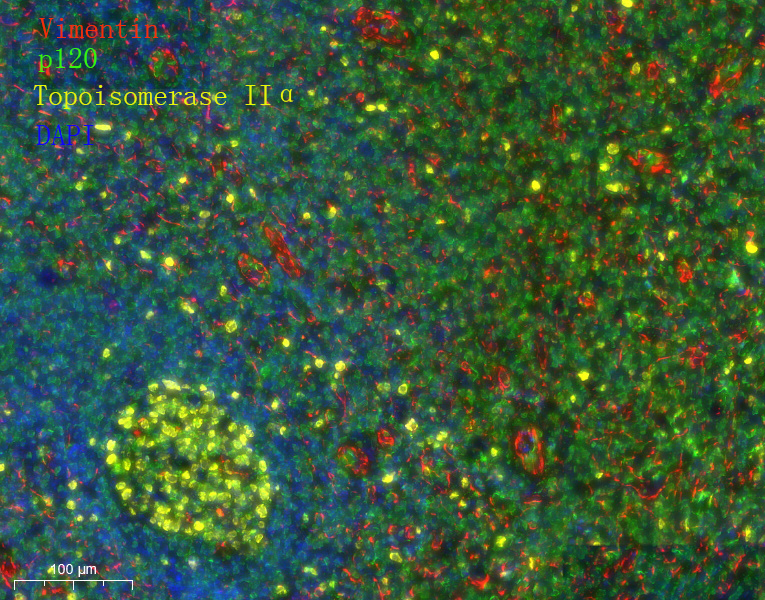 |
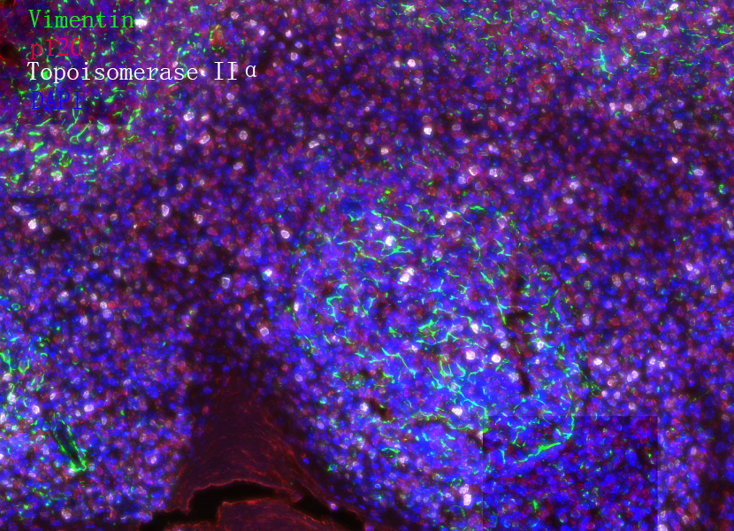
Fluorescence multiplex immunohistochemical analysis of Human tonsil tissue .
The section was incubated in 3 rounds of staining; sequentially for Anti-Vimentin (1:200), Anti-p120 (1:200), Anti-Topoisomerase IIα (1:200).; each using a separate fluorescent tyramide signal amplification system. EDTA based antigen retrieval (Leica Biosystems BOND® Epitope Retrieval Solution 2, pH 9.0, 20 minutes) was used in between rounds of tyramide signal amplification to remove the antibody from the previous round, to avoid any cross-reactivity. DAPI (dark blue) was used as a nuclear counter stain.
Microscopy and pseudocoloring of individual dyes was performed using a Slideviewer Imaging System (3D histech). |
 |
Fluorescence multiplex immunohistochemical analysis of Human tonsil tissue .
The section was incubated in 3 rounds of staining; sequentially for Anti-Vimentin (1:200), Anti-p120 (1:200), Anti-Topoisomerase IIα (1:200).; each using a separate fluorescent tyramide signal amplification system. EDTA based antigen retrieval (Leica Biosystems BOND® Epitope Retrieval Solution 2, pH 9.0, 20 minutes) was used in between rounds of tyramide signal amplification to remove the antibody from the previous round, to avoid any cross-reactivity. DAPI (dark blue) was used as a nuclear counter stain.
Microscopy and pseudocoloring of individual dyes was performed using a Slideviewer Imaging System (3D histech). |


 收藏
收藏

 电话咨询
电话咨询
 在线咨询
在线咨询
 QQ
QQ
 二维码
二维码
 扫码二维码
扫码二维码