ITA6 (10H16) Mouse Monoclonal Antibody

货号: BD-PB3786
收藏
规格 价格 货期 数量
  • 50μl
    1100.00
    现货
    100μl
    2000.00
    现货

产品基本信息

  • 产品货号 BD-PB3786
  • 别名 Integrin alpha-6, CD49 antigen-like family member F, VLA-6, CD49f, Integrin alpha-6 heavy chain, Integrin alpha-6 light chain, Processed integrin alpha-6, Alpha6p, ITGA6
  • 产品名称 ITA6 (10H16) Mouse Monoclonal Antibody
  • 类别 抗体产品
  • Clonality Monoclonal
  • 推荐应用 WB, IHC-P, IF-ICC, FCM
  • 反应种属 Human, Mouse
  • Human Gene ID NP_000201.2;NP_001073286.1
  • Human Swissprot No. P23229
  • 特异性 ITA6 recombinant protein is used to produce this monoclonal antibody.
  • 稀释度 WB 1:100-16000, IHC-P 1:100-500, IF-ICC 1:10-50, FCM 1:10-50
  • 预测分子量 127kDa
  • 同种型 IgG1,κ
  • 背景介绍 The ITGA6 protein product is the integrin alpha chain alpha 6. Integrins are integral cell-surface proteins composed of an alpha chain and a beta chain. A given chain may combine with multiple partners resulting in different integrins. For example, alpha 6 may combine with beta 4 in the integrin referred to as TSP180, or with beta 1 in the integrin VLA-6. Integrins are known to participate in cell adhesion as well as cell-surface mediated signalling. Two transcript variants encoding different isoforms have been found for this gene.
  • 组织表达 Integrin alpha-6/beta-4 is predominantly expressed by epithelia. Isoforms containing segment X1 are ubiquitously expressed. Isoforms containing segment X1X2 are expressed in heart, kidney, placenta, colon, duodenum, myoblasts and myotubes, and in a limited number of cell lines; they are always coexpressed with the ubiquitous isoform containing segment X1. In some tissues (e.g Salivary gland), isoforms containing cytoplasmic segment A and isoforms containing segment B are detected while in others, only isoforms containing one cytoplasmic segment are found (segment A in epidermis and segment B in kidney). Processed integrin alpha-6: Expressed at low levels in normal prostate tissue with elevated levels in prostate cancer tissue (at protein level) (PubMed:15023541)
  • 细胞定位 Cell membrane; Single-pass type I membrane protein. Cell membrane; Lipid-anchor

图片

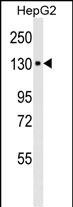
Western blot analysis of anti-ITA6 in HepG2 cell line lysates (35μg/lane). ITA6 (arrow) was detected using the purified Mab.(8μg/ml)
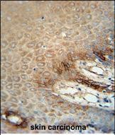
ITA6 Monoclonal Antibody immunohistochemistry analysis in formalin fixed and paraffin embedded human skin carcinoma followed by peroxidase conjugation of the secondary antibody and DAB staining. This data demonstrates the use of the ITA6 Monoclonal Antibody for immunohistochemistry. Clinical relevance has not been evaluated.
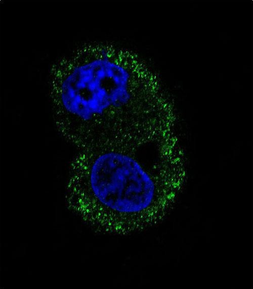
Confocal immunofluorescent analysis of ITA6 Antibody with HepG2 cell followed by Alexa Fluor® 488-conjugated goat anti-mouse lgG (green). DAPI was used to stain the cell nuclear (blue).
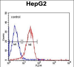
ITA6 Monoclonal Antibody flow cytometric analysis of HepG2 cells (right histogram) compared to a negative control cell (left histogram).PE-conjugated goat-anti-mouse secondary antibodies were used for the analysis.

产品详情

文献引用 (0)

产品问答

内容已折叠

相关产品

内容已折叠

最新浏览的产品

评论内容:
上传图片:
商品评价:
提交评论
« »